-prepared
by Meredith Peach
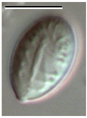
a
cnida from
Catostylus
mosaicus
(click to enlarge)
Cnidae
Many people have been stung by jellyfish medusae at some time in their lives. The stings are caused by tiny capsules called cnidae (singular: cnida), which contain a coiled tubule and venom. In medusae, most cnidae are located in and around the tentacles and/or oral arms, although some cnidae are also found in the bell. Upon contact with another animal, the tubule is discharged in a harpoon-like fashion, either entangling or piercing the skin of the animal it contacts. In this way, jellyfish capture and immobilise zooplankton prey, and in some cases they may also discourage predators. Cnidae are contained within specialised cells called cnidocytes, that bear sensory hairs (cilia) which may be instrumental in discharging the cnidae. Chemical cues also seem to be involved.
Cnidae are divided into two main categories; nematocysts and spirocysts (Mariscal 1974). Scyphozoans have only nematocysts, but within this category there is considerable variety. Over thirty morphological types of nematocysts have been identified, and various systems of nomenclature based on morphology have been proposed over the past few decades (for a succinct summary of this confusing nomenclature, see Östman 2000).
The array of nematocyst types and sizes, called the cnidom or cnidome, can be useful in the identification and taxonomy of scyphozoans. Fully describing the cnidome of a species requires extensive sampling, however, as the cnidome can vary according to location within an individual jellyfish. For example, the tentacles may have a different array of nematocyst types or sizes to the bell. The cnidome may also vary among conspecific individuals of different sizes, and at different life history stages. Comprehensive cnidomes have not yet been described for the majority of scyphozoan species.
|
-prepared
by Meredith Peach
|
||||
|
a
cnida from |
||||